Click image to see more details
-

-
-
-
-
+6
Product Info Summary
| SKU: | A04934-2 |
|---|---|
| Size: | 100 μg/vial |
| Reactive Species: | Human, Mouse, Rat |
| Host: | Rabbit |
| Application: | ELISA, IF, IHC, ICC, WB |
Customers Who Bought This Also Bought


Product info
Product Name
Anti-ORP150/HYOU1 Antibody Picoband®
View all ORP150/HSP12A Antibodies
SKU/Catalog Number
A04934-2
Size
100 μg/vial
Form
Lyophilized
Description
Boster Bio Anti-ORP150/HYOU1 Antibody Picoband® catalog # A04934-2. Tested in ELISA, IF, IHC, ICC, WB applications. This antibody reacts with Human, Mouse, Rat. The brand Picoband indicates this is a premium antibody that guarantees superior quality, high affinity, and strong signals with minimal background in Western blot applications. Only our best-performing antibodies are designated as Picoband, ensuring unmatched performance.
Storage & Handling
At -20°C for one year from date of receipt. After reconstitution, at 4°C for one month. It can also be aliquotted and stored frozen at -20°C for six months. Avoid repeated freezing and thawing.
Cite This Product
Anti-ORP150/HYOU1 Antibody Picoband® (Boster Biological Technology, Pleasanton CA, USA, Catalog # A04934-2)
Host
Rabbit
Contents
Each vial contains 4 mg Trehalose, 0.9 mg NaCl, 0.2 mg Na2HPO4.
Clonality
Polyclonal
Isotype
Rabbit IgG
Immunogen
E.coli-derived human ORP150/HYOU1 recombinant protein (Position: K46-K913).
*Blocking peptide can be purchased. Costs vary based on immunogen length. Contact us for pricing.
Cross-reactivity
No cross-reactivity with other proteins.
Reactive Species
A04934-2 is reactive to HYOU1 in Human, Mouse, Rat
Reconstitution
Adding 0.2 ml of distilled water will yield a concentration of 500 μg/ml.
Observed Molecular Weight
150 kDa
Calculated molecular weight
56903 MW
Background of ORP150/HSP12A
Hypoxia up-regulated protein 1 is a protein that in humans is encoded by the HYOU1 gene. The protein encoded by this gene belongs to the heat shock protein 70 family. This gene uses alternative transcription start sites. A cis-acting segment found in the 5' UTR is involved in stress-dependent induction, resulting in the accumulation of this protein in the endoplasmic reticulum (ER) under hypoxic conditions. The protein encoded by this gene is thought to play an important role in protein folding and secretion in the ER. Since suppression of the protein is associated with accelerated apoptosis, it is also suggested to have an important cytoprotective role in hypoxia-induced cellular perturbation. This protein has been shown to be up-regulated in tumors, especially in breast tumors, and thus it is associated with tumor invasiveness. This gene also has an alternative translation initiation site, resulting in a protein that lacks the N-terminal signal peptide. This signal peptide-lacking protein, which is only 3 amino acids shorter than the mature protein in the ER, is thought to have a housekeeping function in the cytosol. In rat, this protein localizes to both the ER by a carboxy-terminal peptide sequence and to mitochondria by an amino-terminal targeting signal. Alternative splicing results in multiple transcript variants.
Antibody Validation
Boster validates all antibodies on WB, IHC, ICC, Immunofluorescence, and ELISA with known positive control and negative samples to ensure specificity and high affinity, including thorough antibody incubations.
Application & Images
Applications
A04934-2 is guaranteed for ELISA, IF, IHC, ICC, WB Boster Guarantee
Assay Dilutions Recommendation
The recommendations below provide a starting point for assay optimization. The actual working concentration varies and should be decided by the user.
Western blot, 0.1-0.25 μg/ml, Human, Mouse, Rat
Immunohistochemistry(Paraffin-embedded Section), 1-2 μg/ml, Human, Mouse, Rat
Immunocytochemistry/Immunofluorescence, 5 μg/ml, Human
ELISA, 0.1-0.5 μg/ml, Human
Positive Control
WB: human MCF-7 whole cell, human HEK293 whole cell, human SH-SY5Y whole cell, human Hela whole cell, rat liver tissue, rat pancreas tissue, mouse liver tissue, mouse pancreas tissue
IHC: human gall bladder adenosquamous carcinoma tissue, human hyroid papillary carcinoma tissue, human laryngeal squamous cell carcinoma tissue, human liver cancer tissue, human lung cancer tissue, mouse colon tissue, mouse brain tissue, rat brain tissue
ICC/IF: CACO-2 cell
Validation Images & Assay Conditions
Click image to see more details
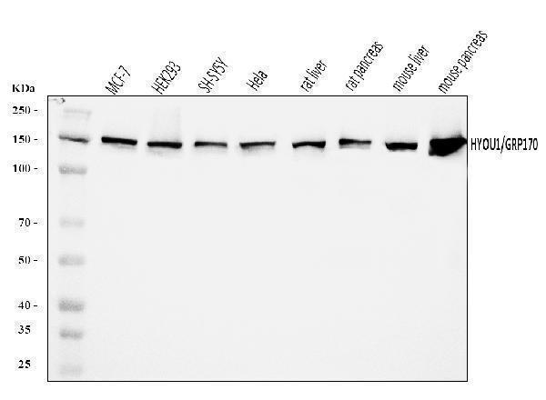
Figure 1. Western blot analysis of ORP150/HYOU1 using anti-ORP150/HYOU1 antibody (A04934-2).
Electrophoresis was performed on a 5-20% SDS-PAGE gel at 70V (Stacking gel) / 90V (Resolving gel) for 2-3 hours. The sample well of each lane was loaded with 30 ug of sample under reducing conditions.
Lane 1: human MCF-7 whole cell lysates,
Lane 2: human HEK293 whole cell lysates,
Lane 3: human SH-SY5Y whole cell lysates,
Lane 4: human Hela whole cell lysates,
Lane 5: rat liver tissue lysates,
Lane 6: rat pancreas tissue lysates,
Lane 7: mouse liver tissue lysates,
Lane 8: mouse pancreas tissue lysates.
After electrophoresis, proteins were transferred to a nitrocellulose membrane at 150 mA for 50-90 minutes. Blocked the membrane with 5% non-fat milk/TBS for 1.5 hour at RT. The membrane was incubated with rabbit anti-ORP150/HYOU1 antigen affinity purified polyclonal antibody (Catalog # A04934-2) at 0.25 μg/mL overnight at 4°C, then washed with TBS-0.1%Tween 3 times with 5 minutes each and probed with a goat anti-rabbit IgG-HRP secondary antibody at a dilution of 1:5000 for 1.5 hour at RT. The signal is developed using an Enhanced Chemiluminescent detection (ECL) kit (Catalog # EK1002) with Tanon 5200 system. A specific band was detected for ORP150/HYOU1 at approximately 150 kDa. The expected band size for ORP150/HYOU1 is at 150 kDa.

Click image to see more details
Figure 2. IHC analysis of ORP150/HYOU1 using anti-ORP150/HYOU1 antibody (A04934-2).
ORP150/HYOU1 was detected in a paraffin-embedded section of human gall bladder adenosquamous carcinoma tissue. Heat mediated antigen retrieval was performed in EDTA buffer (pH 8.0, epitope retrieval solution). The tissue section was blocked with 10% goat serum. The tissue section was then incubated with 2 μg/ml rabbit anti-ORP150/HYOU1 Antibody (A04934-2) overnight at 4°C. Biotinylated goat anti-rabbit IgG was used as secondary antibody and incubated for 30 minutes at 37°C. The tissue section was developed using Strepavidin-Biotin-Complex (SABC) (Catalog # SA1022) with DAB as the chromogen.

Click image to see more details
Figure 3. IHC analysis of ORP150/HYOU1 using anti-ORP150/HYOU1 antibody (A04934-2).
ORP150/HYOU1 was detected in a paraffin-embedded section of human hyroid papillary carcinoma tissue. Heat mediated antigen retrieval was performed in EDTA buffer (pH 8.0, epitope retrieval solution). The tissue section was blocked with 10% goat serum. The tissue section was then incubated with 2 μg/ml rabbit anti-ORP150/HYOU1 Antibody (A04934-2) overnight at 4°C. Biotinylated goat anti-rabbit IgG was used as secondary antibody and incubated for 30 minutes at 37°C. The tissue section was developed using Strepavidin-Biotin-Complex (SABC) (Catalog # SA1022) with DAB as the chromogen.

Click image to see more details
Figure 4. IHC analysis of ORP150/HYOU1 using anti-ORP150/HYOU1 antibody (A04934-2).
ORP150/HYOU1 was detected in a paraffin-embedded section of human laryngeal squamous cell carcinoma tissue. Heat mediated antigen retrieval was performed in EDTA buffer (pH 8.0, epitope retrieval solution). The tissue section was blocked with 10% goat serum. The tissue section was then incubated with 2 μg/ml rabbit anti-ORP150/HYOU1 Antibody (A04934-2) overnight at 4°C. Biotinylated goat anti-rabbit IgG was used as secondary antibody and incubated for 30 minutes at 37°C. The tissue section was developed using Strepavidin-Biotin-Complex (SABC) (Catalog # SA1022) with DAB as the chromogen.

Click image to see more details
Figure 5. IHC analysis of ORP150/HYOU1 using anti-ORP150/HYOU1 antibody (A04934-2).
ORP150/HYOU1 was detected in a paraffin-embedded section of human liver cancer tissue. Heat mediated antigen retrieval was performed in EDTA buffer (pH 8.0, epitope retrieval solution). The tissue section was blocked with 10% goat serum. The tissue section was then incubated with 2 μg/ml rabbit anti-ORP150/HYOU1 Antibody (A04934-2) overnight at 4°C. Biotinylated goat anti-rabbit IgG was used as secondary antibody and incubated for 30 minutes at 37°C. The tissue section was developed using Strepavidin-Biotin-Complex (SABC) (Catalog # SA1022) with DAB as the chromogen.

Click image to see more details
Figure 6. IHC analysis of ORP150/HYOU1 using anti-ORP150/HYOU1 antibody (A04934-2).
ORP150/HYOU1 was detected in a paraffin-embedded section of human lung cancer tissue. Heat mediated antigen retrieval was performed in EDTA buffer (pH 8.0, epitope retrieval solution). The tissue section was blocked with 10% goat serum. The tissue section was then incubated with 2 μg/ml rabbit anti-ORP150/HYOU1 Antibody (A04934-2) overnight at 4°C. Biotinylated goat anti-rabbit IgG was used as secondary antibody and incubated for 30 minutes at 37°C. The tissue section was developed using Strepavidin-Biotin-Complex (SABC) (Catalog # SA1022) with DAB as the chromogen.

Click image to see more details
Figure 7. IHC analysis of ORP150/HYOU1 using anti-ORP150/HYOU1 antibody (A04934-2).
ORP150/HYOU1 was detected in a paraffin-embedded section of mouse colon tissue. Heat mediated antigen retrieval was performed in EDTA buffer (pH 8.0, epitope retrieval solution). The tissue section was blocked with 10% goat serum. The tissue section was then incubated with 2 μg/ml rabbit anti-ORP150/HYOU1 Antibody (A04934-2) overnight at 4°C. Biotinylated goat anti-rabbit IgG was used as secondary antibody and incubated for 30 minutes at 37°C. The tissue section was developed using Strepavidin-Biotin-Complex (SABC) (Catalog # SA1022) with DAB as the chromogen.

Click image to see more details
Figure 8. IHC analysis of ORP150/HYOU1 using anti-ORP150/HYOU1 antibody (A04934-2).
ORP150/HYOU1 was detected in a paraffin-embedded section of mouse brain tissue. Heat mediated antigen retrieval was performed in EDTA buffer (pH 8.0, epitope retrieval solution). The tissue section was blocked with 10% goat serum. The tissue section was then incubated with 2 μg/ml rabbit anti-ORP150/HYOU1 Antibody (A04934-2) overnight at 4°C. Biotinylated goat anti-rabbit IgG was used as secondary antibody and incubated for 30 minutes at 37°C. The tissue section was developed using Strepavidin-Biotin-Complex (SABC) (Catalog # SA1022) with DAB as the chromogen.

Click image to see more details
Figure 9. IHC analysis of ORP150/HYOU1 using anti-ORP150/HYOU1 antibody (A04934-2).
ORP150/HYOU1 was detected in a paraffin-embedded section of rat brain tissue. Heat mediated antigen retrieval was performed in EDTA buffer (pH 8.0, epitope retrieval solution). The tissue section was blocked with 10% goat serum. The tissue section was then incubated with 2 μg/ml rabbit anti-ORP150/HYOU1 Antibody (A04934-2) overnight at 4°C. Biotinylated goat anti-rabbit IgG was used as secondary antibody and incubated for 30 minutes at 37°C. The tissue section was developed using Strepavidin-Biotin-Complex (SABC) (Catalog # SA1022) with DAB as the chromogen.

Click image to see more details
Figure 10. IF analysis of ORP150/HYOU1 using anti-ORP150/HYOU1 antibody (A04934-2).
ORP150/HYOU1 was detected in an immunocytochemical section of CACO-2 cells. Enzyme antigen retrieval was performed using IHC enzyme antigen retrieval reagent (AR0022) for 15 mins. The cells were blocked with 10% goat serum. And then incubated with 5 μg/mL rabbit anti-ORP150/HYOU1 Antibody (A04934-2) overnight at 4°C. DyLight®594 Conjugated Goat Anti-Rabbit IgG (BA1142) was used as secondary antibody at 1:100 dilution and incubated for 30 minutes at 37°C. The section was counterstained with DAPI. Visualize using a fluorescence microscope and filter sets appropriate for the label used.
Protein Target Info & Infographic
Gene/Protein Information For HYOU1 (Source: Uniprot.org, NCBI)
Gene Name
HYOU1
Full Name
Hypoxia up-regulated protein 1
Weight
56903 MW
Superfamily
heat shock protein 70 family
Alternative Names
Meiotic recombination protein REC8 homolog; Cohesin Rec8p; REC8; REC8L1 HYOU1 GRP-170, Grp170, HSP12A, IMD59, ORP-150, ORP150 hypoxia up-regulated 1 hypoxia up-regulated protein 1|150 kDa oxygen-regulated protein|170 kDa glucose-regulated protein|epididymis secretory sperm binding protein|oxygen regulated protein (150kD)
*If product is indicated to react with multiple species, protein info is based on the gene entry specified above in "Species".For more info on HYOU1, check out the HYOU1 Infographic

We have 30,000+ of these available, one for each gene! Check them out.
In this infographic, you will see the following information for HYOU1: database IDs, superfamily, protein function, synonyms, molecular weight, chromosomal locations, tissues of expression, subcellular locations, post-translational modifications, and related diseases, research areas & pathways. If you want to see more information included, or would like to contribute to it and be acknowledged, please contact [email protected].
Specific Publications For Anti-ORP150/HYOU1 Antibody Picoband® (A04934-2)
Hello CJ!
No publications found for A04934-2
*Do you have publications using this product? Share with us and receive a reward. Ask us for more details.
Recommended Resources
Here are featured tools and databases that you might find useful.
- Boster's Pathways Library
- Protein Databases
- Bioscience Research Protocol Resources
- Data Processing & Analysis Software
- Photo Editing Software
- Scientific Literature Resources
- Research Paper Management Tools
- Molecular Biology Software
- Primer Design Tools
- Bioinformatics Tools
- Phylogenetic Tree Analysis
Customer Reviews
Have you used Anti-ORP150/HYOU1 Antibody Picoband®?
Submit a review and receive an Amazon gift card.
- $30 for a review with an image
0 Reviews For Anti-ORP150/HYOU1 Antibody Picoband®
Customer Q&As
Have a question?
Find answers in Q&As, reviews.
Can't find your answer?
Submit your question
