Click image to see more details
-

-
-
-
-
+1
Product Info Summary
| SKU: | A00076-2 |
|---|---|
| Size: | 100 μg/vial |
| Reactive Species: | Human, Rat |
| Host: | Rabbit |
| Application: | ELISA, Flow Cytometry, IHC, WB |
Customers Who Bought This Also Bought


Product info
Product Name
Anti-LDL Receptor/LDLR Antibody Picoband®
SKU/Catalog Number
A00076-2
Size
100 μg/vial
Form
Lyophilized
Description
Boster Bio Anti-LDL Receptor/LDLR Antibody Picoband® catalog # A00076-2. Tested in ELISA, Flow Cytometry, IHC, WB applications. This antibody reacts with Human, Rat. The brand Picoband indicates this is a premium antibody that guarantees superior quality, high affinity, and strong signals with minimal background in Western blot applications. Only our best-performing antibodies are designated as Picoband, ensuring unmatched performance.
Storage & Handling
Store at -20˚C for one year from date of receipt. After reconstitution, at 4˚C for one month. It can also be aliquotted and stored frozen at -20˚C for six months. Avoid repeated freeze-thaw cycles.
Cite This Product
Anti-LDL Receptor/LDLR Antibody Picoband® (Boster Biological Technology, Pleasanton CA, USA, Catalog # A00076-2)
Host
Rabbit
Contents
Each vial contains 4mg Trehalose, 0.9mg NaCl, 0.2mg Na2HPO4, 0.05mg NaN3.
Clonality
Polyclonal
Isotype
Rabbit IgG
Immunogen
E.coli-derived human LDL Receptor/LDLR recombinant protein (Position: Q35-D843).
*Blocking peptide can be purchased. Costs vary based on immunogen length. Contact us for pricing.
Cross-reactivity
No cross-reactivity with other proteins.
Reactive Species
A00076-2 is reactive to LDLR in Human, Rat
Reconstitution
Add 0.2ml of distilled water will yield a concentration of 500ug/ml.
Observed Molecular Weight
130 kDa
Calculated molecular weight
95.376kDa
Background of LDLR
In humans, the LDL receptor protein is encoded by the LDLR gene on chromosome 19. It is mapped to 19p13.2. The low density lipoprotein receptor (LDLR) gene family consists of cell surface proteins involved in receptor-mediated endocytosis of specific ligands. Low density lipoprotein (LDL) is normally bound at the cell membrane and taken into the cell ending up in lysosomes where the protein is degraded and the cholesterol is made available for repression of microsomal enzyme 3-hydroxy-3-methylglutaryl coenzyme A (HMG CoA) reductase, the rate-limiting step in cholesterol synthesis. At the same time, a reciprocal stimulation of cholesterol ester synthesis takes place. Mutations in this gene cause the autosomal dominant disorder, familial hypercholesterolemia. Alternate splicing results in multiple transcript variants.
Antibody Validation
Boster validates all antibodies on WB, IHC, ICC, Immunofluorescence, and ELISA with known positive control and negative samples to ensure specificity and high affinity, including thorough antibody incubations.
Application & Images
Applications
A00076-2 is guaranteed for ELISA, Flow Cytometry, IHC, WB Boster Guarantee
Assay Dilutions Recommendation
The recommendations below provide a starting point for assay optimization. The actual working concentration varies and should be decided by the user.
Western blot, 0.25-0.5μg/ml, Human, Rat
Immunohistochemistry (Paraffin-embedded Section), 0.5-1μg/ml, Human
Flow Cytometry (Fixed), 1-3μg/1x106 cells, Human, Rat
Direct ELISA, 0.1-0.5μg/ml, Human
Positive Control
WB: human Hela whole cell, human Raji whole cell, human U-87MG whole cell, rat liver tissue, rat lung tissue, rat kidney tissue, rat RH35 whole cell
IHC: human liver cancer tissue, human rectal cancer tissue
FCM: A431 cell, NRK cell
Validation Images & Assay Conditions
Click image to see more details
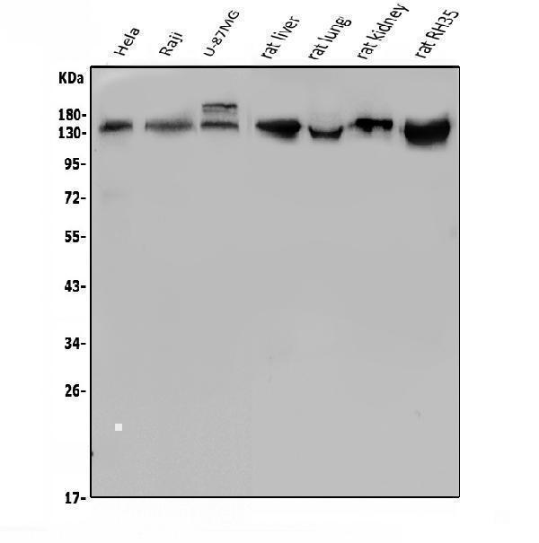
Figure 1. Western blot analysis of LDLR using anti-LDLR antibody (A00076-2).
Electrophoresis was performed on a 5-20% SDS-PAGE gel at 70V (Stacking gel) / 90V (Resolving gel) for 2-3 hours. The sample well of each lane was loaded with 50ug of sample under reducing conditions.
Lane 1: human Hela whole cell lysates,
Lane 2: human Raji whole cell lysates,
Lane 3: human U-87MG whole cell lysates,
Lane 4: rat liver tissue lysates,
Lane 5: rat lung tissue lysates,
Lane 6: rat kidney tissue lysates,
Lane 7: rat RH35 whole cell lysates.
After Electrophoresis, proteins were transferred to a Nitrocellulose membrane at 150mA for 50-90 minutes. Blocked the membrane with 5% Non-fat Milk/ TBS for 1.5 hour at RT. The membrane was incubated with rabbit anti-LDLR antigen affinity purified polyclonal antibody (Catalog # A00076-2) at 0.5 μg/mL overnight at 4°C, then washed with TBS-0.1%Tween 3 times with 5 minutes each and probed with a goat anti-rabbit IgG-HRP secondary antibody at a dilution of 1:5000 for 1.5 hour at RT. The signal is developed using an Enhanced Chemiluminescent detection (ECL) kit (Catalog # EK1002) with Tanon 5200 system. A specific band was detected for LDLR at approximately 130KD. The expected band size for LDLR is at 95KD.

Click image to see more details
Figure 2. IHC analysis of LDLR using anti-LDLR antibody (A00076-2).
LDLR was detected in paraffin-embedded section of human liver cancer tissue. Heat mediated antigen retrieval was performed in EDTA buffer (pH8.0, epitope retrieval solution). The tissue section was blocked with 10% goat serum. The tissue section was then incubated with 1μg/ml rabbit anti-LDLR Antibody (A00076-2) overnight at 4°C. Biotinylated goat anti-rabbit IgG was used as secondary antibody and incubated for 30 minutes at 37°C. The tissue section was developed using Strepavidin-Biotin-Complex (SABC) (Catalog # SA1022) with DAB as the chromogen.

Click image to see more details
Figure 3. IHC analysis of LDLR using anti-LDLR antibody (A00076-2).
LDLR was detected in paraffin-embedded section of human rectal cancer tissue. Heat mediated antigen retrieval was performed in EDTA buffer (pH8.0, epitope retrieval solution). The tissue section was blocked with 10% goat serum. The tissue section was then incubated with 1μg/ml rabbit anti-LDLR Antibody (A00076-2) overnight at 4°C. Biotinylated goat anti-rabbit IgG was used as secondary antibody and incubated for 30 minutes at 37°C. The tissue section was developed using Strepavidin-Biotin-Complex (SABC) (Catalog # SA1022) with DAB as the chromogen.

Click image to see more details
Figure 4. Flow Cytometry analysis of A431 cells using anti-LDLR antibody (A00076-2).
Overlay histogram showing A431 cells stained with A00076-2 (Blue line). To facilitate intracellular staining, cells were fixed with 4% paraformaldehyde and permeabilized with permeabilization buffer. The cells were blocked with 10% normal goat serum. And then incubated with rabbit anti-LDLR Antibody (A00076-2, 1μg/1x106 cells) for 30 min at 20°C. DyLight®488 conjugated goat anti-rabbit IgG (BA1127, 5-10μg/1x106 cells) was used as secondary antibody for 30 minutes at 20°C. Isotype control antibody (Green line) was rabbit IgG (1μg/1x106) used under the same conditions. Unlabelled sample without incubation with primary antibody and secondary antibody (Red line) was used as a blank control.

Click image to see more details
Figure 5. Flow Cytometry analysis of NRK cells using anti-LDLR antibody (A00076-2).
Overlay histogram showing NRK cells stained with A00076-2 (Blue line). To facilitate intracellular staining, cells were fixed with 4% paraformaldehyde and permeabilized with permeabilization buffer. The cells were blocked with 10% normal goat serum. And then incubated with rabbit anti-LDLR Antibody (A00076-2, 1μg/1x106 cells) for 30 min at 20°C. DyLight®488 conjugated goat anti-rabbit IgG (BA1127, 5-10μg/1x106 cells) was used as secondary antibody for 30 minutes at 20°C. Isotype control antibody (Green line) was rabbit IgG (1μg/1x106) used under the same conditions. Unlabelled sample without incubation with primary antibody and secondary antibody (Red line) was used as a blank control.
Protein Target Info & Infographic
Gene/Protein Information For LDLR (Source: Uniprot.org, NCBI)
Gene Name
LDLR
Full Name
Low-density lipoprotein receptor
Weight
95.376kDa
Superfamily
LDLR family
Alternative Names
Low-density lipoprotein receptor; LDL receptor; LDLR LDLR FH, FHC, FHCL1, LDLCQ2 low density lipoprotein receptor low-density lipoprotein receptor|LDL receptor|low-density lipoprotein receptor class A domain-containing protein 3
*If product is indicated to react with multiple species, protein info is based on the gene entry specified above in "Species".For more info on LDLR, check out the LDLR Infographic

We have 30,000+ of these available, one for each gene! Check them out.
In this infographic, you will see the following information for LDLR: database IDs, superfamily, protein function, synonyms, molecular weight, chromosomal locations, tissues of expression, subcellular locations, post-translational modifications, and related diseases, research areas & pathways. If you want to see more information included, or would like to contribute to it and be acknowledged, please contact [email protected].
Specific Publications For Anti-LDL Receptor/LDLR Antibody Picoband® (A00076-2)
Hello CJ!
A00076-2 has been cited in 3 publications:
*The publications in this section are manually curated by our staff scientists. They may differ from Bioz's machine gathered results. Both are accurate. If you find a publication citing this product but is missing from this list, please let us know we will issue you a thank-you coupon.
Difference in LDL Receptor Feedback Regulation in Macrophages and Vascular Smooth Muscle Cells: Foam Cell Transformation Under Inflammatory Stress
Inflammatory stress increases unmodified LDL uptake via LDL receptor: an alternative pathway for macrophage foam-cell formation
Zha K,Ye Q.Golgi a-mannosidase II mediates the formation of vascular smooth muscle foam cells under inflammatory stress.Folia Histochem Cytobiol.2021; 59(2):134-143.doi:10.5603/FHC.a2021.0015.Epub 2021 Jun 21.PMID:34151999.
Species: Human
A00076-2 usage in article: APP:WB, SAMPLE:VSMCS, DILUTION:1:1500
Recommended Resources
Here are featured tools and databases that you might find useful.
- Boster's Pathways Library
- Protein Databases
- Bioscience Research Protocol Resources
- Data Processing & Analysis Software
- Photo Editing Software
- Scientific Literature Resources
- Research Paper Management Tools
- Molecular Biology Software
- Primer Design Tools
- Bioinformatics Tools
- Phylogenetic Tree Analysis
Customer Reviews
Have you used Anti-LDL Receptor/LDLR Antibody Picoband®?
Submit a review and receive an Amazon gift card.
- $30 for a review with an image
0 Reviews For Anti-LDL Receptor/LDLR Antibody Picoband®
Customer Q&As
Have a question?
Find answers in Q&As, reviews.
Can't find your answer?
Submit your question
